Histopathology of enamel caries from birefringence: the surface layer according to Darling (1958)
HISTOPATHOLOGY OF ENAMEL CARIES FROM BIREFRINGENCE: THE SURFACE LAYER ACCORDING TO A.I. DARLING (Br Dent J, 105: 119-135, 1958)
1) Why polarized light microscopy is so special for enamel?
Because:
Light is retarded by interacting with electric fields in heterogeneous materials, and in the extreme case light is absorbed. Thus, before absorption occurs contrast can be obtained by retardance. PLM is more sensitive for contrasting normal from carious enamel than absorption-based techniques.
2) Image of enamel under polarized light microscopy is due to the observed birefringence (BRobs)
This was already known in the early 20th century, and researchers looked for a mathematical explanation of enamel BRobs.
3) Form birefringence
•Given by the Wiener (Abh. Sächs. Akad. Wiss. Math. Phys., 32: 509–604., 1912) equation for inhomogeneous materials (including enamel)
V1 = mineral volume;
V2 = non-mineral volume (1-V1);
n1 = mineral phase refractive index (1.62 for enamel);
n2 = non-mineral phase refractive index
For a more recent reference on the Wiener equation see Oldenbourg & Ruiz (Biophys J, 56: 195-205, 1989).
Contribution from A.G. Darling (Br Dent J, 105: 119-135, 1958)
In order to interpret enamel BRobs, Darling recognized that form birefringence could be described by the Wiener equation, but there was no equation for enamel intrinsic birefringence. Darling performed a laboratory experiment to measure the intrinsic birefringence of enamel and found a negative value of – 0.003.
Assumption used by Darling:
- Enamel intrinsic birefringence is a fixed value not influenced by mineral volume.
Problem with the assumption
- As early as in the 19th century it was recognized that intrinsic birefringence is proportional to the material’s volume.
Enamel form birefringence according to Darling (Br Dent J, 105: 119-135, 1958):
Model's implications
- All pores are completely filled by the immersion medium;
- This was Darling’s approximation in the late 50’s, but it was already known that enamel pores were not filled by a single phase only. As no one had a better model, it was accepted.
4 ) The mathematical approach of Darling
BRobs = BRintr + BRform = -0.003 + Wiener equation

The methodology for measuring enamel mineral volume was not well established in the 50’s. Darling needed to solve this problem because mineral volume is required in the Weiner equation.

The solution was to plot BRobs as a function of mineral volume and then to match experimental BRobs with calculated BRobs.

As the degree of BRform was considered to be always at 100% (not less, not more), all changes in enamel BRobs in a single immersion medium were attributed to changes in mineral volume.
Interpretation of enamel birefringence
- Model of Darling (1958): immersion in water.
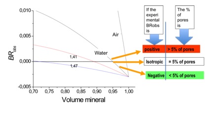
Plot of the BRobs as a function of the mineral volume according to the model of Darling (1958). All BRobs values for mineral volumes < 0.7 (70%) are positive.
The interpretation is:
- As, from theory (see figure plot above), negative birefringence in water occurs for mineral volume > 95% (5% of pores), the negative BRobs of enamel experimentally detected in the laboratory can be interpreted as presenting less than 5% of pores (the same as normal enamel).
- Thus, the negative BRobs of the surface layer of enamel caries presents porosity of less than 5%. As this is the same behavior of normal enamel, the surface layer can be regarded as a relatively intact layer.
Problems with the interpretation
- The mineral volume used in calculations is not experimentally determined; it is a theoretical construct, meaning that less than 5% of pores does not actually mean a mineral volume of 95%;
- porosity is measured with an ordinal (not scale) variable;
- enamel pores are not completely filled by a single component (i.e., water in this case); when air dried, enamel losses only part of its water content, so that air and/or Thoulet's solutions cannot completely replace water in enamel;
- from general theory, organic matter can be expected to alter BRobs;
- the intrinsic birefringence is a function of the actual mineral volume; the model of intrinsic birefringence of Darling assumes that it does not vary with mineral volume;
- normal enamel is variable, deceasing from the occlusal to the cervical region and from the surface to the enamel-dentin junction. Darling considered it as a fixed value in the equation of intrinsic birefringence.
Implications
- It was accepted a reasonable description of the surface layer of enamel caries;
- Surface layer was a result of remineralization, following Darling’s assumptions;
- Increase in the thickness of the surface layer is interpreted as remineralization;
- As microradiographs qualitatively revealed radiolucency in the body of the lesion only (more than 5% of pores, according to Darling), many researchers in the field of Cariology still think that microradiography cannot detect mineral loss less than 5%.
Experimental data from independent reports supporting Darling (1958) -------------------
- No experimental data supported the interpretation that the surface layer has less than 5% of pores;
- Bergman & Ove (J Dent Res, 45:1477, 1966), using microradiography, reported mineral volumes of the surface layer ranging from 77.9 to 85.8 % (based on a mineral density of 3.15 gcm-3); the corresponding mineral loss (relative to normal enamel) ranged from 0.3% to 11.1% (mean of 6%)
Experimental data from independent reports not supporting Darling (1958) ------------------
- the mineral volume of the surface layer has been reported in some cases to be lower than that of both the body of the lesion and normal enamel (Medeiros et al., J Microsc, 246:177, 2012; Dijkman et al., Caries Res, 20:202, 1986; Mellberg et al., J Dent Res, 67:1461,1988; Ogaard et al., 20:270, 1986).
There are open questions to be answered ...
1) Why polarized light microscopy is so special for enamel?
Because:
Light is retarded by interacting with electric fields in heterogeneous materials, and in the extreme case light is absorbed. Thus, before absorption occurs contrast can be obtained by retardance. PLM is more sensitive for contrasting normal from carious enamel than absorption-based techniques.
2) Image of enamel under polarized light microscopy is due to the observed birefringence (BRobs)
This was already known in the early 20th century, and researchers looked for a mathematical explanation of enamel BRobs.
3) Form birefringence
•Given by the Wiener (Abh. Sächs. Akad. Wiss. Math. Phys., 32: 509–604., 1912) equation for inhomogeneous materials (including enamel)
V1 = mineral volume;
V2 = non-mineral volume (1-V1);
n1 = mineral phase refractive index (1.62 for enamel);
n2 = non-mineral phase refractive index
For a more recent reference on the Wiener equation see Oldenbourg & Ruiz (Biophys J, 56: 195-205, 1989).
Contribution from A.G. Darling (Br Dent J, 105: 119-135, 1958)
In order to interpret enamel BRobs, Darling recognized that form birefringence could be described by the Wiener equation, but there was no equation for enamel intrinsic birefringence. Darling performed a laboratory experiment to measure the intrinsic birefringence of enamel and found a negative value of – 0.003.
Assumption used by Darling:
- Enamel intrinsic birefringence is a fixed value not influenced by mineral volume.
Problem with the assumption
- As early as in the 19th century it was recognized that intrinsic birefringence is proportional to the material’s volume.
Enamel form birefringence according to Darling (Br Dent J, 105: 119-135, 1958):
Model's implications
- All pores are completely filled by the immersion medium;
- This was Darling’s approximation in the late 50’s, but it was already known that enamel pores were not filled by a single phase only. As no one had a better model, it was accepted.
4 ) The mathematical approach of Darling
BRobs = BRintr + BRform = -0.003 + Wiener equation

The methodology for measuring enamel mineral volume was not well established in the 50’s. Darling needed to solve this problem because mineral volume is required in the Weiner equation.

The solution was to plot BRobs as a function of mineral volume and then to match experimental BRobs with calculated BRobs.

As the degree of BRform was considered to be always at 100% (not less, not more), all changes in enamel BRobs in a single immersion medium were attributed to changes in mineral volume.
Interpretation of enamel birefringence
- Model of Darling (1958): immersion in water.
Image of ground section of natural enamel caries under water immersion in polarized light microscopy (dark background; without Red I filter).
Plot of the BRobs as a function of the mineral volume according to the model of Darling (1958). All BRobs values for mineral volumes < 0.7 (70%) are positive.
The interpretation is:
- As, from theory (see figure plot above), negative birefringence in water occurs for mineral volume > 95% (5% of pores), the negative BRobs of enamel experimentally detected in the laboratory can be interpreted as presenting less than 5% of pores (the same as normal enamel).
- Thus, the negative BRobs of the surface layer of enamel caries presents porosity of less than 5%. As this is the same behavior of normal enamel, the surface layer can be regarded as a relatively intact layer.
Problems with the interpretation
- The mineral volume used in calculations is not experimentally determined; it is a theoretical construct, meaning that less than 5% of pores does not actually mean a mineral volume of 95%;
- porosity is measured with an ordinal (not scale) variable;
- enamel pores are not completely filled by a single component (i.e., water in this case); when air dried, enamel losses only part of its water content, so that air and/or Thoulet's solutions cannot completely replace water in enamel;
- from general theory, organic matter can be expected to alter BRobs;
- the intrinsic birefringence is a function of the actual mineral volume; the model of intrinsic birefringence of Darling assumes that it does not vary with mineral volume;
- normal enamel is variable, deceasing from the occlusal to the cervical region and from the surface to the enamel-dentin junction. Darling considered it as a fixed value in the equation of intrinsic birefringence.
Implications
- It was accepted a reasonable description of the surface layer of enamel caries;
- Surface layer was a result of remineralization, following Darling’s assumptions;
- Increase in the thickness of the surface layer is interpreted as remineralization;
- As microradiographs qualitatively revealed radiolucency in the body of the lesion only (more than 5% of pores, according to Darling), many researchers in the field of Cariology still think that microradiography cannot detect mineral loss less than 5%.
Experimental data from independent reports supporting Darling (1958) -------------------
- No experimental data supported the interpretation that the surface layer has less than 5% of pores;
- Bergman & Ove (J Dent Res, 45:1477, 1966), using microradiography, reported mineral volumes of the surface layer ranging from 77.9 to 85.8 % (based on a mineral density of 3.15 gcm-3); the corresponding mineral loss (relative to normal enamel) ranged from 0.3% to 11.1% (mean of 6%)
Experimental data from independent reports not supporting Darling (1958) ------------------
- the mineral volume of the surface layer has been reported in some cases to be lower than that of both the body of the lesion and normal enamel (Medeiros et al., J Microsc, 246:177, 2012; Dijkman et al., Caries Res, 20:202, 1986; Mellberg et al., J Dent Res, 67:1461,1988; Ogaard et al., 20:270, 1986).
There are open questions to be answered ...


0 Comments:
Post a Comment
<< Home